Archive for the ‘Uncategorized’ Category

FINAL BLOG ENTRY

April 25, 2008

For my final blog entry I chose to write on the phenomenon of synesthesia which I saw as the most interesting and surprising condition that I encountered during the semester.  I had before seen cartoons and other such things on television portraying the senses and symptoms produced by synesthesia but never realized that the condition actually existed.  Before having relevant knowledge of the condition, the symptoms produced by the synesthete I would believe could only be explained through the use of hallucinogenic drugs.  Thus, the fact that the condition is actually produced by the body’s own sensory system and cognitive faculty without the use of such stimulants makes synesthesia a truly remarkable phenomenon. As a result, I did some research on the condition with much of the information provided coming from the book Synesthesia: A Union of the Senses by Richard Cytowic.


Synesthesia derives its name from the Ancient Greek (syn), which means “with,” and (aisthēsis), meaning “sensation.”  The condition is neurologically-based in which stimulation of one sensory or cognitive pathway causes an involuntary, separate response in another sensory or cognitive pathway.  Historically, although synesthesia has been known for about 300 years, there has been little research on the condition because the sciences of psychology and neurology have only begun to bloom in their own respects within the last 70 or so years.  Only in recent years has cognition been associated brain function.  Currently there are 54 known types of synesthesia, each shown in the chart below which was given in a lecture given by Professor Leanne Boucher.

One type of synesthesia, called color graphemic synesthesia, is characterized by people seeing colors when looking at letters or numbers.  In another subform of graphemic synesthesia, ordinal linguistic personification, numbers, days, or months produce personalities.  It is noted that there are many variations in the senses produced.  For example, one synesthete may “have a highly restricted form of colored hearing, for

example, in which only a particular voice or particular kind of music

will elicit photisms. The opposite extreme is the pentamodal patient:

stimulation of one sense causes synesthesia in the remaining four” (Cytowic, Section 2). However, just by viewing the graph above one can determine the characteristic of each type of synesthesia and also determine the percentage of the prevalence that each form occurs.


Cytowic notes that in most instances the features of synesthesia have been present in a patient as far back as the patient can remember.  Even as young children these synesthetes are able to realize that their perceptual world is markedly different from that of others.  Also, many times these people keep their special symptoms to themselves believing that if they shared the story of their condition with others that they would be teased because others would not believe them. Synesthesia also brings with it psychological influences that can greatly affect the personalities of its subjects.  However, it is also noted that although the condition brings with it many burdens, most synesthetes would not trade their abnormal senses for those of normal individuals.  They, in turn, believe their perceptions to be real as their symptoms remain with them their entire lives.


In his book, Cytowic also gives personal accounts of the experiences of a number of synesthetes.  Below is two such examples given from patient MN and patient OM.


Patient MN: “I remember most accurately scents. We were preparing to move into the house I grew up in. I remember at age 2 my father was on a ladder painting the

left side of the wall. The paint smelled blue, although he was painting it white. I

remember to this day thinking why the paint was white, when it smelled blue” (Cytowic, Section 2.1).


Patient OM: “Colors are very important to me because I have a gift—it’s not my fault, it’s just how I am—whenever I hear music, or even if I read music, I see colors” (Cytowic, Section 2.1).


Cytowic writes that when he encountered his first synesthete patient in 1979 that the interest in the condition was almost nonexistent.  At this time he was criticized by his colleagues telling him that he was wasting his time and that he would likely ruin his career by focusing on such a bizarre condition.  Since then he acknowledges that scientists in 13 countries have begun to show interest and investigate different aspects of synesthesia.  Along with the increased interest in perceptual synesthesia, there has also begun a growing interest in metaphoric, or historical, synesthesia which can be seen by the number of research and encyclopedia pertaining to the subject.  Currently, there are only three scientists predominately involved in the research of the condition.  “They are Simon Baron-Cohen, an experimental psychologist in England; Hinderk Emrich, a philosopher turned psychiatrist and head of the clinical psychiatry department at the medical school in Hannover, Germany; and [Cytowic], an American neurologist and neuropsychologist (with a smidgen of British training)” (Cytowic, 3.1). Cytowic further notes that the three involved in the research of synesthesia each approach the condition in ways that are relevant to their basic fields of study.


Cytowic also lays out the diagnostic criteria that characterize idiopathic synesthesia which are discussed in his book.  He also notes that these criteria help to distinguish idiopathic synesthesia from acquired synesthesia including drug-induced synesthesia, epileptic synesthesia, and that acquired through brain lesions.  The criteria for this condition include:

  1. “Synesthesia is involuntary but elicited”
  2. ‘Synesthesia is spatially extended”
  3. “Synesthetic percepts are consistent and discrete”
  4. “Synesthesia is memorable”
  5. “Synesthesia is emotional”


The theories of the mechanism of synesthesia are grouped into the following categories:

1. Undifferentiated neuronal activity: sensory incontinence analogous

to synkinesis in infants

2. Linkage theories

a. Neural specificity

b. Polymodal combination

c. Vestigial connections, persisting from birth

3. Abstractions theories

a. Cognitive mediation

b. Aristotelian common sense (Cytowic, Section 3.4)


For the category of undifferentiated neuronal activity, previous beliefs theorized synesthesia’s cause to be linked to an immature nervous system and compared the condition to ordinary syn-kinesis, which is joined movements, seen in all infants.  During syn-kinesis, movements in one area extend to other muscle groups.  It is believed that this movement is not fixed until corticospinal and cerebellar motor pathways have matured and achieved thorough myelination.  Cytowic states that this theory is labeled the degeneracy theory or compensation theory and further suggests that “synesthesia is a form of atavism or sensory incontinence” (Cytowic, Section 3.4.1).  However, he also states that this theory is no longer seriously considered and proceeds to discuss the similarity of synesthesia to the gustofacial reflex, which is a “well-differentiated motor reaction of the facial muscles to taste stimuli” (Cytowic, Section 3.4.1).  Comparisons of synesthesia and the gustofacial reflex suggest that the neural mechanism of synesthesia lies above the level of the brainstem. 


The linkage theories of synesthesia suggest that there is a difference in the circuitry of synthetic brains compared to those on nonsynesthetes.  Cytowic notes the significance of the synesthesia that occasionally occurs with LSD users and other anti-serotonergics since the hallucinations that are perceived by these individuals are assisted via sensory input from other modalities.  Using LSD studies, it is also proposed that emotional meaning might be an appropriate linkage to the perceptions of synesthetes.  Cytowic further notes that synesthetic patients are normally capable of making intermodal associations but that the difference lies with an additional binds to lead to perceptual experiences that are not additive but, instead, integrated.  He also states that Emrich labels this hyperbinding.  Citing a study by Maurer (1993), it also believed that if all neonates are synesthetic but lose their condition by 6 months of age then vestigial remnants could help explain synesthesia in these individuals in adulthood.  Furthermore, citing the study of Paulesu and colleagues (1995), it is noted that cerebral metabolism is disturbed in synesthetic patients from what is expected in normal, nonsynesthetic individuals.  Lastly, Cytowic admits that the question of “What is the nature of the proposed connection?” still remains.  While Baren-Cohen propose corticocortical connections, Cytowic and Emrich support a vertical, lower-level linkage via corticolimbic projections.


The two main versions of the abstraction theory, cognitive mediation and Aristotelian common sense, both theorize a that there is a “filtering out” of certain sense elements that leave one with “an abstract emotional or an abstract perceptual residue that serves

as a synesthetic mediator” (Cytowic, Section 3.4.3).  It is noted that in research done by Marks (1978), he believes synesthesia to be basically perceptual in nature and that such perception without language can still have meaning.  Cytowic states that Marks has demonstrated that “even in cross-modal perception by nonsynesthetic persons, language only modulates but does not wholly mask or replace the underlying and prior sensory relations. In nonsynesthetic children, cross-modal similarities are stronger perceptually than verbally” (Cytowic, 3.4.3). 

The emotional tone theory, which is still part of the abstraction theory, asserts that since synesthesia and its stimulus are common emotionally to the synesthete then the sensation produced should be one composed of responses from many of the senses and not just restricted to that of only one.  However, this is not commonly seen is the responses seen in synesthetes.

Aristotelian common senses propose that the sensations produced through synesthesia are mediated through language.  Thus, this theory suggests that synesthesia is more language-based and an more centered around metaphoric language than actual sensations.  However, Cytowic claims that such “linguistic theories” are not correct.  Finally, Cytowic states due to Aristotle’s common senses theory, research for the last 100 years has concentrated on shared meanings as the link to synesthesia and proposed that the condition took place at the highest levels of abstract processing in the central nervous system.


The information above is a just a dent in that provided on the many aspects of synesthesia.  Synesthesia: A Union of the Senses supplies a great deal of more information that I have not listed and there are numerous other books in the Vanderbilt libraries as well as on the internet that can help better explain the phenomenon of synesthesia.

This is a painting by Jane Mackay titled “Tchaikovsky’s First Piano Concerto” which was created to portray a synesthetic composition that connect the senses of sight and sound.

(http://images.google.com/imgres?imgurl=http://research.yale.edu/ysm/images/77.4/articles-synesthesia-brain.jpg&imgrefurl=http://research.yale.edu/ysm/article.jsp%3FarticleID%3D83&h=300&w=400&sz=36&hl=en&start=1&tbnid=8Cvv0cA4s5ZtVM:&tbnh=93&tbnw=124&prev=/images%3Fq%3Dsynesthesia%26gbv%3D2%26hl%3Den)


This is to show an example of what a synesthete will experience.  When looking at the picture above most people will see many 5’s and if they look longer will notice that some of the 5’s have been reversed to make 2’s.  For someone with number-color synesthesia, they will immediately see a triangle of 2’s.  The triangle would stand out because the 5’s and 2’s would both appear in different colors.  Below is a portrayal of the picture to both a nonsynesthete and a synesthete.

(http://images.google.com/imgres?imgurl=http://www.youramazingbrain.org/images/brainchanges/synesthesia.gif&imgrefurl=http:  //www.youramazingbrain.org/brainchanges/synesthesia.htm&h=320&w=320&sz=9&hl=en&start=4&tbnid=9l1yxQf-hBgGfM:&tbnh=118&tbnw=118&prev=/images%3Fq%3Dsynesthesia%26gbv%3D2%26hl%3Den)


A picture portraying what someone with color-number synesthesia may see when looking at a set of numbers.

(http://images.google.com/imgres?imgurl=http://www.elchill.com/pictures/color_number_synesthesia.jpg&imgrefurl=http:   //www.elchill.com/smec/&h=283&w=544&sz=29&hl=en&start=5&tbnid=PLf-n_0HabFXnM:&tbnh=69&tbnw=133&prev=/images%3Fq%3Dsynesthesia%26gbv%3D2%26hl%3Den)


WORKS CITED:

Cytowic, Richard E. Synesthesia: a Union of the Senses. 2nd ed. Cambridge, Mass.: MIT P, 2002.


http://en.wikipedia.org/wiki/Synesthesia


Randolph, Blake, and Sekuler Robert. Perception. 5th ed. Boston: McGraw Hill, 2006. 269-271.


The Aperture Problem

April 18, 2008

4. What is the aperture problem? 


In motion detection, each motion sensor or directional selectivity (DS) neuron in the visual system is sensitive or responsive only to events that take place within the small part of its own receptive field.  Since each neuron can only respond to stimuli within such a small area of it receptive field, it is as if each neuron is looking through a small aperture.  Because of this minimal window through which the neuron is able to “see,” motion direction of an object can often be ambiguous.  Such ambiguity means that a DS neuron will often produce identical responses for a number of different forms having different orientations traveling at different speeds.  An example of the aperture problem is shown below.  Whether the rectangular structure is moved diagonally, horizontally, or vertically, the image will be identical when viewed through the aperture in all three instances.  This produces ambiguity as to the actual direction, orientation, and speed of the object.


Since these individual neurons of the visual system are only sensitive to motion that occurs within their small receptive field, each producing the aperture problem, we require input from many neurons to fix this problem.  The approximations from the responses of many individual neurons are combined to create a comprehensive motion estimate.  This sort of global motion processing is believed to occur in the area V5/MT of the visual cortex.  The textbook explains that motions of different objects can be either correlated, in which they all move in the same direction, or uncorrelated in which the motion is random.  The function of the MT is to discriminate between correlated and uncorrelated motion in order to determine an object’s direction.  It was also noted that correlations as small as 3 to 5 percent are strong enough to discriminate a motion’s direction. 

Additionally, here is a link to a website showing two clips that can better display the aperture problem. http://www.psico.univ.trieste.it/labs/perclab/integration/english_version/aperture.php3


Citations:

http://en.wikipedia.org/wiki/Motion_perception#The_aperture_problem

Randolph, Blake, and Sekuler Robert. Perception. 5th ed. Boston: McGraw Hill, 2006. 338-345.


Additive and Subtractive Colors Systems

April 12, 2008

2.  What is the difference between adding and subtracting colors?  What does that even mean? 

                                       Figure: Subtractive Coloring

Subtracting colors are caused by a mixture of colors, from either paints, inks, or almost anything within the natural environment, absorbing certain wavelengths of light while subtracting others. Paints, inks, and other such colored things contain pigments that selectively absorb certain wavelengths while reflecting others; therefore, subtracting certain wavelengths from our vision.  The color that we perceive and see is based on which parts of the electromagnetic spectrum that are reflected.  As an example, red paint absorbs shorter wavelengths while reflecting longer wavelengths producing the perceptual color of red that we see.  When mixing one subtractive color with another it further limits the reflected light thereby producing a darker color.  For instance, mixing the three secondary colors magenta, yellow, and cyan produces the color black.  Furthermore, the subtractive color system originates from white light, which is a blend of all the colors in the visual spectrum.  The different paints and other objects or substances then subtract certain wavelengths from this light producing a certain color that we perceive. 


                          Figure: Additive Coloring

While subtractive colors are often seen in nature, additive colors are rarely seen naturally and are often for purposes of entertainment or research.  James Clerk Maxwell is given credit for first showing the concept of additive colors.  He projected the colors of red, green, and blue producing a full color image.  Additive colors, different from subtractive colors, involve light emitted from a light source or some other sort of illuminant thereby forming perceived colors through emission rather than through reflection and also through the process of adding wavelengths rather than subtracting them.  This process usually involves a combination or mixture of the primary colors red, blue, and green to produce other colors often producing secondary colors such as magenta, cyan, and yellow.  Contrary to subtractive colors in which mixing the three secondary colors magenta, cyan, an yellow to produce black, additive colors instead uses a mixture of the three primary colors red, green, and blue to produce white.  Furthermore, additive colors due not coincide with the normal color mixtures that we are used to in our everyday lives such as mixing blue and yellow to produce green.  An example of an additive mixture is combining red and green to produce yellow, although not simple combination can be used to produce green.  Moreover, additive colors are produced from the way our eyes detect different colors instead of being the property of light such as with subtractive colors.  The properties of our nervous system allow us to perceive a wide range of colors through the additive coloring system.  For example, with subtractive colors, our nervous system perceives the color yellow through its reflected wavelength of about 5700-580 nm.  The perception of the additive color yellow is on the other hand a mixture of red and green light.


                     Figure: Absorption Spectra of Cone and Rod Cells (http://upload.wikimedia.org/wikipedia/commons/thumb/6/65/Cone-response.svg/410px-Cone-response.svg.png)                    

          Lastly, whether with additive or subtractive colors, our perception of colors is accomplished by matching with our trichromatic color vision.  Our trichromatic color vision is caused by the three types of cones cells in the body, S, M, and L, each corresponding and being sensitive to a certain range of wavelengths.  These three types of cones  respond both to the light and intensity of the light that reaches them through the retina.  Through having to depend on both light and intensity, the brain often needs input from a combination of two types of cones to be able to properly perceive color. 

(Citations:       http://en.wikipedia.org/wiki/Additive_color

                        http://en.wikipedia.org/wiki/Subtractive_color

                        http://en.wikipedia.org/wiki/Trichromatic

Randolph, Blake, and Sekuler Robert. Perception. 5th ed. Boston: McGraw Hill, 2006. 243-244.)


Results of an Under- or Abnormally Developed Visual System

April 3, 2008

 Strabismic Amblyopia

 Figure: Strabismic Amblyopia

2.  What are the behavioral consequences of an under-developed or abnormally developed visual system?

During childhood, the visual system grows and develops at the same time that the brain matures, a process that takes about ten years.  While babies are capable of seeing, their vision is poor at first and limited to a vision of about 20/1500.  This is due to the brain not maturing yet to the level where it is able to learn to process the visual information that it receives.  As the visual system further develops, vision will become clearer and will develop so that the child will eventually be able to see the slightest details of an image.  However, the visual system of some children becomes underdeveloped or abnormally developed causing them to have visual disorders.  Oftentimes these visual disorders are related to brain disorders that are also present within the child.  Some types of vision problems include cortical blindness, delayed visual maturation, optic nerve hypoplasia, optic nerve atrophy, cranial neuropathies, along with many others.           

One commonly seen disorder from an underdeveloped visual system is amblyopia, also known as “lazy eye,” in which the visual function of one eye is underdeveloped while the vision of the other eye functions normally.  Any factor that prevents or inhibits clear vision during childhood can promote amblyopia.  Some of the chief causes of amblyopia, which are abnormal developments in themselves, are strabismus (misalignment of the eyes), unequal focus due to an asymmetrical refractive error, and cloudiness in normally clear eye tissues such as corneal opacities and cataracts which prevent correct focus of light in the eye.             

Another visual defect that was already mentioned above is strabismus in which the eyes develop misaligned and point in different directions.  This disorder is often most common among children but is also seen in about 2 percent of the adult population also.  Because of the eye misalignment, the brain receives two different visual messages.  In children, the brain will often disregard or not put as much emphasis on the image sent by the diverging eye while still processing highly detailed visual information from the straight eye. This improper functioning results in amblyopia causing one eye to become “lazy.”  Strabismus is also commonly associated with defective or absent binocular vision, which is characterized by reduced 3-D vision, resulting in impaired depth perception.  As the visual system further develops into adulthood, strabismus may cause double vision, eye strain, discomfort in reading, and headaches. Adult who develop strabismus with no prior history of childhood strabismus may have medical or neurological causes of the defect which may be due to diabetes, thyroid disease, myasthenia gravis, brain tumor, or stroke.

                Although I have only mentioned two defects caused by an underdeveloped or abnormally developed visual system there are a number of other disorders.  Many minor defects such as myopia or hyperopia can lead to blurry and unclear vision but can be corrected through corrective lenses or refractive surgery.  Other disorders such as glaucoma or retinopathy of prematurity can cause blindness while others such as retinoblastoma, a form of cancer, can eventually lead to death.  The large number of defects and disorders caused by an under- or abnormally developed nervous system are also accompanied with a vast number of consequences.  Heredity, genetics, nutrition, environmental toxins, stress, problems at birth, or insufficient sensory stimulation can all be blamed as causes for these disorders or defects and the results that they produce.


(Citation: http://www.bpei.med.miami.edu/site/disease/disease_pediatric.asp)


Oblique Effect and the Organization of Visual Information

March 23, 2008

 fMRI and behavioral measurements of an oblique effect in human striate cortex

fMRI and behavioral measurements of an oblique effect in human striate cortex                                                                                                   (Reference: http://www.nature.com/neuro/journal/v3/n6/fig_tab/nn0600_535_F1.html)   

3. How can we use the oblique effect to study the organization of visual information? 

The oblique effect is a visual defect in which vertical and horizontal lines are more easily detected or identified quicker than lines that are oriented obliquely.  The oblique effect is seen in most humans; however, some experience the reverse of the oblique effect and still others fail to experience it at all.  Those who do not experience the oblique effect correctly usually have some degree of astigmatism.  In certain cases in which the oblique orientation is preferred for vision, this preference still continues even after the astigmatism is correct.  As a result, all orientations appear equally clear on the retina, a condition known as meridional amblyopia.  This shows that even prior to correction the brain was oriented in a way that it preferred oblique over horizontal or vertical lines.  Animal studies have shown that in the brains of animals that prefer oblique lines, that the brain was altered in a way to favor such a preference.  The cortical cells in these animals favor orientations that are centered around the one that is the clearly focused, while fewer cells respond to the unclear astigmatic orientation.  This shows that biased individual experience can change the orientation preferences of cortical cells and it is further theorized that people with meridional amblyopia have less cortical cells tuned to the formerly blurred orientation. 

            Furthermore, there are two theories to explain why vertical and horizontal orientation is more highly favored.  The first theory believes that we have developed this preference because of the carpenter environment we live in in which vertical and horizontal contours are abundant.  Because of this predisposed visual exposure, orientation preferences among cortical cells in the brain are influenced.  Another theory assumes that the neural basis for vertical and horizontal preference can be attributed to genetic factors that prefer the developmental of cortical cells to favor such orientations.

Off-Center Retinal Ganglion Cells

March 16, 2008

2. What is an OFF-center ganglion cell receptive field?  Why is it organized they way it is?

 The way in which a ganglion receptive field is arranged provides a way of detecting contrast and is used for detecting the edges of objects.  The receptive field is a template that determines what pattern of light will best stimulate the neuron.  Receptive fields are arranged with a central disk, labeled the centre, and a concentric ring that surrounds the centre, labeled the surround.  The off-center ganglion receptive field is the area surrounding the center, or the surround.  Each region of a ganglion receptive field responds oppositely to light, e.g., light hitting the centre of the receptive field might increase the firing rate of a particular ganglion cell while light hitting the surrounding (off-center) receptor field would decrease the rate of firing and vice versa.  Stimulation of the surround in an off-center ganglion cell’s receptive field would produce depolarization and a high frequency firing rate of the ganglion cell while stimulation of the centre would produce the opposite effect, hyperpolarization and a low frequency firing rate.  Stimulation of both areas simultaneously would produce a mild firing rate of both areas due to mutual inhibition from the on and off-center regions. When photoreceptors of the off-center cell are stimulated, they respond by inhibiting the release of glutamate.  At the synapses with ganglion cells, glutamate acts as an excitatory neurotransmitter by opening channels that depolarize the cell.  The inhibition of glutamate caused a decrease in the firing rate of the on-center cell.  The ganglion cell receptive field is organized the way it is because photoreceptors are able to make synapses with both on-center and off-center cells and, in turn, contains synapses in which glutamate has an inhibitory effect and ones in which it has an excitatory effect.  This organization provides information not only about whether photoreceptors are firing but also the rates at which different cells (on- and off-center) are firing which provides information about contrast.  Furthermore, the size of the receptive field controls the spatial frequency of the information.  Small receptive field are stimulated by high spatial frequencies showing fine detail while large receptive fields are stimulated by low spatial frequencies showing rough detail.  These cells provide information about the discontinuities in the delivery of light reaching the retina; this usually provides information about the edges of objects.

Rotating Snake’s Illusion

March 9, 2008

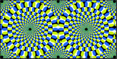
Rotating Snake’s Illusion

2. Investigate your favorite optical illusion and find out why it works the way it does. 

The Rotating Snakes Illusion is a peripheral drift illusion which refers to a motion illusion produced by the appearance of a sawtooth luminance grating in the visual periphery.  This illusion of motion is produced by static repeated asymmetric patterns (RAPs) which can cause many people’s visual systems to detect motion when there actually is none.  With many visual illusions such as the Rotating Snakes Illusion it is believed that small involuntary eye movements while looking at the pattern play a significant role in producing such illusions.  Furthermore, it is believed that such illusions mainly result from fast and slow changes over time in the neuronal representation of contrast-driven RAPs or luminance-driven RAPs.  With contrast-driven RAPs such as the Rotating Snakes Illusion, the temporal phase advance in the neuronal response at high contrast can explain the initial, fast motion after each fixation change.  The basis behind this explanation is that motion detectors neglect to compensate for the process of neuronal encoding.  However, the theory is very complicated and detailed in trying to explain the neurological basis behind the Rotating Snakes Illusion.  For detailed information on the illusion here is a link to the article Illusory motion from change over time in the response to contrast and luminance:

http://journalofvision.org/5/11/10/article.aspx

Interaural Cues

February 23, 2008

1.  We use interaural cues to locate acoustic stimuli, but this information isn’t always so clear.  How might one disambiguate information presented within these “cones of confusion”?  And why are we not always confused about a sound’s location? 

The duplex theory of sound localization for explaining sound localization attributes this ability to two different interaural cues which include interaural time differences and interaural level differences.  The interaural time difference is a binaural auditory cue which is the minute time difference between when sound from a single source reaches the near ear and when it reaches the far ear.  This slight time difference provides an essential cue to direction. The interaural level difference is the reduction in loudness that occurs when a sound reaches the far ear.  With the head being in the way, it makes it difficult to calculate the correct interaural time difference and the interaural level difference since there may not always be a direct path for a sound to make it from a source to the far ear. This produces an intensity difference in the sound from one ear to the other.  However, if a sound comes from anywhere in the median plane, which could cause the sound to reach both ears at the same time and with the same intensity, then the person will perceive the sound as being somewhere in the plane but often cannot tell where in the plane it actually occurs resulting in what is called the “plane of confusion.”  In much the same way, the person experiences a “cone of confusion” for sounds when sounds come from one side of the median plane.  In this situation the person is able to tell which side the sound comes from but is unable to differentiate among sounds that come from the many points on the surface of the cone.  The confusion comes from the fact that all the points on the cone are equal distances from one ear than from the other which results in the same difference in stimuli at both ears.  One way to overcome this is to simply tilt one’s head. This creates different temporal patterns of stimulation arriving at the two ears allowing people to differentiate among sounds that that would otherwise be unable to detect as far as direction is concerned.

Skittle Experiment

February 9, 2008

3. Re-do the skittle experiment with some friends (block the nose, try to guess which skittle was presented) and perform a signal detection analysis on the data. Some ways to do this (1) test 2 flavors only (grape and lemon, for example) and ask your friend to identify if the skittle was one of the flavors (grape, for example), (2) do what is suggested in (1) twice, once with flavors that are similar in taste (i.e., green and yellow) and once with flavors that are different in taste (i.e., purple and orange).


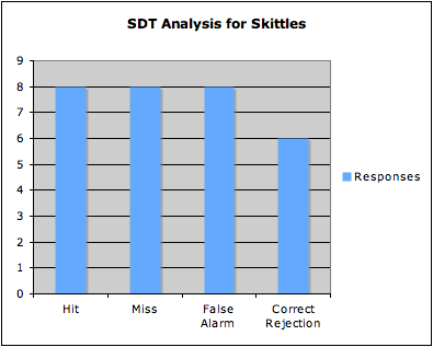
After looking at the options for the blog entries this week, Alok and I decided to re-do the skittle experiment with a group of friends and perform a signal detection analysis with our results.  For the experiment we used three different friends and gave them ten trials each.  For the first friend we used yellow and orange skittles by randomly giving him either yellow or orange on each trial in which he was supposed to say whether the skittle was orange or not.  For the other two friends, we performed the same experiment using purple and red skittles in which they were supposed to say whether the skittle was purple or not.  The complete results from the experiment gave 8 hits, 8 misses, 8 false alarms, and 6 correct rejections.  Below is a bar graph showing the results from the experiment.

Results


The Fallen Angel of the Senses

February 1, 2008

3. Smell is often described as the “fallen angel” of the senses. Why is that?

Helen Keller once said that smell is the “fallen angel” of the senses and, in many respects, she is correct.  For humans, we often neglect our sense of smell and rarely use it actively.  Instead, we have the tendency to rely on our other senses such as sight, hearing, and touch to guide us through our everyday lives.  For me, personally, this makes sense as I realize that smell is likely my weakest sense.  As I go throughout my day, my sense of smell is often out of my own consciousness until I am around or walk by something that has a strong odor and even in these instances I oftentimes cannot put a label on certain smells or even identify the object from which the smell comes from. Instead I use my other senses, such as sight, as guidance.  Through evolution and our growing mental capacity that comes with it, we have not had to rely on our sense of smell to the degree that many animals do and are able to use our other senses and our ability to reason to substitute for our sense of smell.

However, although most people take our sense of smell for granted, many companies are taking advantage of the pleasure and enjoyment that smell can bring to us, oftentimes out of our own conscious awareness.  Different fragrances can affect our moods, relieve stress, encourage relaxation and even improve our alertness which has been seen through the practice of aromatherapy. Also, much like we talked about in class on Wednesday, malls will often filter the smell of different foods from the food court into the main areas of the mall to attract people to buy their food products and real estates companies will place coffee and fresh baked cookies or muffins in the homes they are selling to give it a more comfortable, homely feeling to name a few examples.  The consumer often does not realize that they are being “duped” into buying these products through their own sense of smell.  As a result, the fact that smell has been called the “fallen angel” of the senses may not actually be due to our sense of small becoming worse and of less use to us. Instead, it may be related to it being out of our conscious awareness most of the time as we have learned to rely on our other senses in its place and frequently neglect the pleasures that many smells can provide.

A link to a New York Times article on how smell can affect our mood: http://query.nytimes.com/gst/fullpage.html?res=9D0CE7DE1531F934A15752C1A967958260&sec=&spon=&pagewanted=all